Siu-Keung LAM
The pathogenesis of invasive cervical cancer and its premalignant stages (cervical intraepithelial neoplasia CIN, also known as SIL squamous intraepithelial lesion) is multifactorial. There are extensive epidemiological data linking human papillomavirus (HPV) infection with invasive carcinoma of cervix and its precursor CIN. More than 100 types of human HPV have been reported of which 30 can infect the anogenital tract. A majority of the cervical cancer worldwide is associated with HPV-16 and HPV-18 infection which are now recognised as oncogenic viruses. HPV DNA can be detected in over 80% of such tumours. Other co-factors predisposing patients to cervical cancer include early coitus, multiple sexual partners, smoking and prolonged use of oral contraceptive pills.
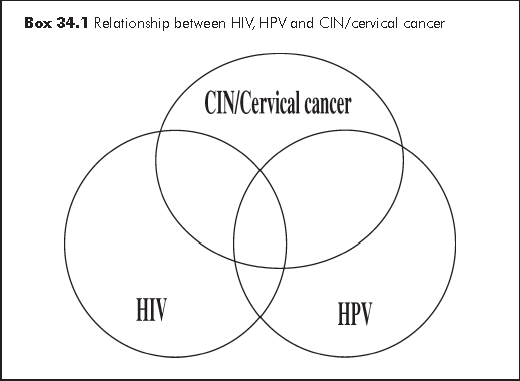
The interplay between HIV infection, HPV infection and CIN/cervical cancer is complex (see Box 34.1). Cervical dysplasia and possibly invasive cervical cancer are more prevalent in HIV-positive women. The latter has a higher rate of HPV infections which are strongly associated with high-grade SIL and invasive cervical cancer.1 Immune suppression is the factor predisposing HIV positive women to HPV infections. CIN is commoner in HIV infected women with a lower CD4 count or AIDS. Sun et al2 have suggested that the presence of immunosuppression shifts the ratio of latent: clinically expressed HPV infection from 8:1 in the general population to 3:1 in HIV-positive women with CD4 >500/μL and to 1:1 when CD4<200/μL. Linking the US AIDS and Cancer Registry, the observed cervical cancer cases in HIV infected women were up to 9 folds higher than the expected number of cases but the likelihood of cervical cancer is not related to the CD4 count.3
For surveillance purpose, invasive cervical cancer has been added to the list of AIDS-defining illnesses in the 1993 revised classification system recommended by the US Centers for Disease Control and Prevention (CDC) and this has also been adopted in Hong Kong. Cervical dysplasia and cervical carcinoma in situ are included as Category B conditions which are attributable to HIV infection or are indicative of the defective cell-mediated immunity. Locally, there have so far not been reports of cervical cancer as a primary AIDS-defining illness.

Invasive cervical cancer carries significant morbidity and mortality. Prevention, early diagnosis and treatment of cervical neoplasia are integral components of management in HIV/AIDS women. Cervical screening for premalignant lesion is the strategy. Compared to one-off screening, periodic regular screening is more effective and therefore advocated. Screening can be considered at two levels: (a) screening for specific oncogenic HPV and (b) screening of early pathological lesions by Pap smear. The benefit of Pap smear screening in HIV infection is well-established, while that of HPV DNA screening either as primary or as adjunct to Pap smear remains to be determined.
After obtaining a complete history of previous cervical diseases, an HIV infected woman should undergo a comprehensive gynaecological examination as baseline, including a pelvic examination and Pap smear as part of the initial evaluation (see Algorithm 34(A)). The wooden Ayres spatula and the glass slide (conventional Pap smear) was the standard in the past. The incidence of unsatisfactory smear especially in the presence of blood and/or infection is high. Currently most centres in Hong Kong use the liquid base cytology which has a lower unsatisfactory rate. HPV DNA test can be performed on the same specimen later if needed.
Pap smear should be repeated in the case of an unsatisfactory specimen. Repeat smears should not be performed till 6-8 weeks have lapsed, to allow time for the scrapped surface to re-epithelialise. It must be noted that most cancers and precancerous lesions arise in the transformation zone. It is essential that the whole transformation zone be sampled in the examination. Most laboratories in Hong Kong report Pap smear results using the Bethesda System 2001:4
(a) specimen adequacy
(b) general categorisation - within normal limits or with specific descriptive diagnosis
(c) descriptive diagnosis - including benign cellular changes, reactive and reparative cellular change
(d) epithelial cell abnormality -
- atypical squamous cells (ASC), which include two categories:
ASC-US (atypical squamous cells of undetermined significance)
ASC-H (atypical squamous cells cannot exclude HGSIL)- low grade SIL (LGSIL), including HPV changes and mild dysplasia/CIN 1
- high grade SIL (HGSIL), including moderate and severe dysplasia, CIN 2, CIN 3, carcinoma in situ
- squamous cell carcinoma
(e) glandular cell abnormalities
(f) non-epithelial malignant neoplasm
Pelvic examination and Pap smear are repeated six months after the baseline.5 If both times are normal, cervical screening is then performed every twelve months alongside with careful vulval, vaginal and anal inspections. There is no specific CD4 threshold under which the frequency of Pap smear would need to be increased, though this may be considered in cases of:5
(a) previous abnormal Pap smear
(b) HPV infection
(c) symptomatic HIV disease
(d) CD4 counts <200/μL
(e) post-treatment for CIN
HPV-DNA has recently been introduced as one form of screening. As there is a high incidence of HPV infection in HIV patient and most HIV patients with abnormal smear will ultimately need colposcopy evaluation (to rule out CIN and cervical cancer), the use of HPV-DNA in the triage for colposcopy is of limited value. On the other hand, despite the higher incidence of cervical abnormality in HIV infected patients, colposcopy is not generally recommended for primary cervical screening6 but indicated when pap smear reveals epithelial cell abnormality (see Algorithm 34(A)).
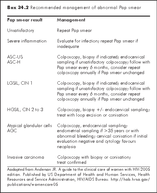
HIV-infected patients with ASC-US or above once on Pap smear should be referred for colposcopy. At a colposcopy clinic, thorough evaluation of the lower genital tract is performed including colposcopy and cervical biopsy at the most suspicious area to rule out CIN and/or malignancy. If there is no HGSIL, patients are followed up regularly in the colposcopy clinic. If HGSIL is found, most patients require large loop excision of transformation zone (LLETZ), to reduce the chance of progression to cervical cancer. This has to be coupled with regular post-treatment cervical smear surveillance. A retrospective review of 13 HIV infected patients with cervical smear abnormality seen in Kwong Wah Hospital showed HPV/cervicitis in 6 patients, CIN 1 in 2 patients, CIN 2 in 4 patients and none has CIN 3 or cancer. Six patients had LLETZ under local anaesthesia in clinic.7 Subsequent to this review, another 22 HIV infected patients were seen in the same clinic and HGSIL was noted in 5 patients. Seven patients required LLETZ. No major postoperative complications nor cervical cancer were noted at follow-up (Lam SK, unpublished data). The role of HAART in preventing CIN recurrence post treatment is still controversial. Box 34.2 shows the recommendation on management of abnormal pap smear by US Health Department.5 The local practice is similar.
When invasive cervical cancer is diagnosed, the patient should be staged properly according to FIGO (International Federation of Obstetrics and Gynaecology) classification and appropriate treatment arranged in oncology centre. For early stages disease (up to Ib), radical hysterectomy should be considered especially in young patient to preserve the ovarian and sexual function. For advanced stages or poor surgical candidates, primary radiotherapy or chemo-radiation can be considered.

Anal HPV-DNA has been reported in up to 76% of HIV positive women. When concurrent anal and cervical HPV data were available, anal HPV was more prevalent in both HIV-infected and high risk HIV-uninfected women.8 Abnormal anal cytology or anal squamous intraepithelial lesion (ASIL or AIN) are reported in up to 26% of HIV-infected women. Risk factors include lower CD4 count, increased HIV viral load, history of receptive anal intercourse and concurrent abnormal cervical cytology.9 Anal smear screening is not recommended as a routine in HIV infected women (and homosexual men). If considered for selected cases, anoscopy and biopsy are needed in patients with abnormal anal smear to rule out AIN and anal cancer.
Sun XW, Kuhn L, Ellerbrock TV, Chiasson MA, Bush TJ, Wright TC Jr. Human papillomavirus infection in women infected with the human immunodeficiency virus. N Engl J Med 1997;337:1343-9.
Sun XW, Ellerbrock TV, Lungu O, Chiasson MA, Bush TJ, Wright TC Jr. Human papillomavirus infection in human immunodeficiency virus-seropositive women. Obstet Gynecol 1995;85(5 Pt 1):680-6.
Mbulaiteye SM, Biggar RJ, Goedert JJ, Engels EA. Immune deficiency and risk for malignancy among persons with AIDS. J Acquir Immune Defic Syndr 2003;32:527-33.
Wright TC Jr, Cox JT, Massad LS, Twiggs LB, Wilkinson EJ; ASCCP-Sponsored Consensus Conference. 2001 Consensus Guidelines for the management of women with cervical cytological abnormalities. JAMA 2002;287:2120-9.
Anderson JR. A guide to the clinical care of women with HIV-2005 edition. Published by US Department of Health and Human Services, Health Resources and Service Administration, HIV/AIDS Bureau. Available from http://hab.hrsa.gov/publications/womencare05.
Anderson JR, Paramsothy P, Heilig C, et al. Accuracy of Papanicolaou test among HIV-infected women. Clin Infect Dis 2006;42:562-8.
Lam SK, Chan KS, Tse HY. Cervical premalignant disease and HIV-experience in a local colposcopy clinic. Hong Kong AIDS Conference Abstract Book 2001 page 238-243.
Palefsky JM, Holly EA, Ralston ML, Da Costa M, Greenblatt RM. Prevalence and risk factors for anal human papillomavirus infection in human immunodeficiency virus (HIV)-positive and high-risk HIV-negative women. J Infect Dis 2001;183:383-91.
Holly EA, Ralston ML, Darragh TM, Greenblatt RM, Jay N, Palefsky JM. Prevalence and risk factors for anal squamous intraepithelial lesions in women. J Natl Cancer Inst 2001;93:843-9.